The Molecular and Biomolecular Technologies team is involved in advanced research that explores and develops novel micro/nanostructured systems targeting emerging technologies, theranostic biomedical applications, biomimetics, molecular and biomolecular sensors, molecular electronics etc.
Initiated / Developed themes
- Molecular recognition and self-association processes, intra and inter-molecular interactions
- Supramolecular structures with controlled architecture and functionality
- Bioinspired intelligent micro/nanostructures with applications in medicine, industry and counter-fitting
- Real-time identification and detection of microorganisms using ultrasensitive spectroscopy techniques
- In silico investigation of molecular systems layed onto metallic surfaces, quantum transport processes, and of biomolecular complexes
- Identification of novel antimicrobial and anticancer peptides
- Transport mechanisms through membrane models, protein channels, biological and solid-state nanotubes/nanopores
- Development of exozome-based innovative platforms for theranostic biomedical applications
Expertise
- Fabrication of molecular structures with controlled architecture and functionality, using of molecular beam epitaxy (MBE) in ultra-high vacuum, and of micro-/nano-structured surfaces, using nano-imprint lithography (NIL), for applications in molecular sensory and molecular electronics
- Nanoscale topographical characterization of surfaces using microscopic scanning techniques (STM and AFM) in ultra-high vacuum
- Microorganisms detection by means of Raman and surface-enhanced Raman spectroscopies (SERS), and their differentiation through multiple chemometric analyses (PCA, LDA, PCA-LDA on vibrational spectra)
- Rational design, generation and characterization of short antimicrobial/anticancer peptides
- Atomic to molecular scale (coarse-grained) molecular modelling and simulation of complex biophysical systems (proteins/short peptides, DNA/RNA, bacterial and mammalian membrane models) by means of molecular dynamics and quantum chemistry
- Development of “tight-binding” and ab-initio models for various molecular systems
- In silico computation of free energy profiles/PMFs for transmembrane molecular transport, (self)association, membrane affinity, by means of equilibrium (Umbrella Sampling), non-equilibrium (FR method), flat-based histrogram ((MW)2-XDOS method), molecular docking (affinities)
- Characterization of biomolecules (with therapeutic/pharmaceutical potential, biomimetical, proteins involved in molecular signalling associated to cancer, oxidative stress/neuro-degenerative diseases) by means of computational simulations (molecular dynamics, molecular docking); biochemical and biophysical methods: SEC-MALS, spectroscopic techniques (UV/Vis, fluorescence, Raman), immunodetection (Western Blot), electrophoresis and gel densitometry; biochemical and microbiological methods: antibacterial activity (MIC/MBS and diffusometric method), testing for cellular viability (IC50), enzymatic activity (TTC), metabolic activity (MTT), membrane integrity (LDH), antioxidant activity (SOD, CAT, GPx, GSH), inflammatory markers
- Bacterial cellular growth; Proteins’ expression in E. coli (cloning in expression vectors, PCR, point mutation genesis, DNA segment assembly cloning); Protein purification (sonication, centrifugation, chromatography techniques); Structural characterization by means of X-ray crystallography of proteins, small molecules, biomolecules
Team Leader
Dr. Ioan TURCU – Scientific Researcher I
Expertise: The effects of strong electric fields on biological cells, Electrofusion of biological cells, Scattering of laser radiation on biological cells; Self-assembly of supramolecular systems, molecular recognition.
Members:
Dr. Sanda BOCA-FARCĂU – Scientific Researcher I
Expertise: Nanoparticles and theranostic nanocompounds, Optical and vibrational (micro)spectroscopy, Nanomedicine, Nanotoxicology.
Dr. Ioana Andreea BREZESTEAN – Scientific Researcher III
Expertise: Nanomaterials, Toxins detections, Raman, SERS, Microscopy, Materials characterization, Uv-Vis, Nanoparticle synthesis.
Dr. Paula BULIERIS – Scientific Researcher III
Expertise: Protein Biochemistry, Structural Biology, Biomolecules characterization by biochemical and biophysical methods.
Dr. Alia COLNIŢĂ – Scientific Researcher
Expertise: Vibrational Spectroscopy, Physical Chemistry, Biomolecular Physics.
Dr. Bogdan Ionuţ COZAR – Scientific Researcher III
Expertise: Molecular and Biomolecular Physics, Theoretical Physics.
Dr. Nicoleta DINA – Scientific Researcher II
Expertise: Vibrational Spectroscopy, Analytical Chemistry, Biomolecular Physics, Theoretical Physics and Statistics, Chemometrics, Computational modelling.
Dr. Alexandra FĂRCAŞ– Scientific Researcher
Expertise: .
Dr. Loránt JÁNOSI – Scientific Researcher I
Expertise: Molecular Modeling, Theoretical and Computational Biophysics, Atomic, Molecular and Chemical Physics, Physical Chemistry.
Dr. Sorin Daniel MARCONI – Scientific Researcher II
Expertise: Solid Physics Physics, Nanomaterials, Thin Films, Molecular Physics.
Cristina NUŢ – Technician
Dr. Anca STOICA – Scientific Researcher
Expertise: Cell and molecular biology, Animal physiology, Biochemistry, Hematology.
Dr. Tiberiu SZŐKE-NAGY – Scientific Researcher
Expertise: Molecular and cellular Biology, Molecular Phylogenetic Analysis, Microbiology.
Dr. Andra-Sorina TĂTAR – Scientific Researcher III
Expertise: Noble metal nanoparticle synthesis, Biocompatibilisation, Biofunctionalisation.
 Bio-inspired intelligent micro/nanostructures with applications in medicine, industry and anti-counterfeiting
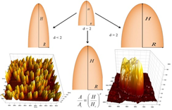
The fabrication of well-defined nanostructures in various materials leads to different and unique properties compared to those of the bulk material, showing high promise in the development of very versatile biosensing platforms. Nanostructured surfaces are of particular interest in the field of medicine, especially in different domains of the healthcare sector: biosensing, drug delivery, biomaterials, implants therapeutics and medical devices and instruments. A particular case of nanostructuring is the formation of Si nanopillars, by using single-step fabrication of homoepitaxial silicon nanocones on Si(111) reconstructed substrates by molecular beam epitaxy technique. [1]. Another focal point deals with the effect of substrate temperature on structural and morphological properties of Au/Si(111) thin films with atomically flat gold terraces used as substrates for self assembled monolayers [2].
[1] A. Colniţă, D. Marconi, R. T. Brătfălean, I. Turcu, Applied Surface Science, Vol. 436, pp. 1163-1172 (2018)
[2] D. Marconi, A. Ungurean, Applied Surface Science, Vol. 288, pp. 166-171 (2014)
Discrimination and Characterization of Gram-Positive Bacteria by Means of Raman Spectroscopy and Principal Component Analysis
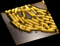
 Raman scattering and its particular effect, surface-enhanced Raman scattering (SERS), are whole-organism fingerprinting spectroscopic techniques that gain more and more popularity in bacterial detection. Two relevant Gram-positive bacteria species, Lactobacillus casei (L. casei) and Listeria monocytogenes (L. monocytogenes) were successfully discriminated based on the joint use of Raman, SERS and Principal Component Analysis (PCA) to their specific spectral data [1]. Furthermore, the biochemical structures of the bacterial cell wall of L. casei and L. monocytogenes were identified by using their Raman and SERS spectral fingerprints. Another recent result has materialized in a patent application submitted to OSIM: A00976/28.11.2018, authors: Dina Nicoleta Elena, Colniţă Alia, Marconi Sorin Daniel, Szöke-Nagy Tiberiu, Gherman Ana-Maria-Raluca, Leopold Nicolae, Ştefancu Andrei, title: Procedeu de detecție prin spectroscopie RAMAN amplificată de suprafață (SERS) într-un sistem de curgere microfluidic utilizând un spot de argint ca substrat amplificator SERS.
Novel Antimicrobial Peptides
One of the main causes leading to antimicrobial (AM) resistance is the very slow progress in the development of new antibacterial drugs. The main challenge for discovering new AM peptides is increasing safety and efficacy of the novel drugs.
Using complementary computational and experimental techniques, we showed that the substitution of tryptophan by histidine in short tryptophan- and arginine-rich peptides proved to strongly modulate the antimicrobial activity, mainly by changing the peptide-to-membrane binding energy [1]. The substitution of arginine has low effect on the antimicrobial efficacy. The presence of histidine residue reduced the cytotoxic and hemolytic activity of the peptides. The peptides’ antimicrobial activity was correlated to the 3D-hydrophobic moment and to a simple structure-based packing parameter. Significance: The short tryptophan-rich peptides’ therapeutic index can be maximized using the histidine substitution to optimize their structure.
Adsorption parameters for organic/magnetic molecules on metallic surfaces
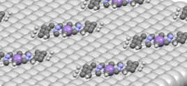
 The characteristics of interaction between transition-metal molecules and metallic surfaces ware detailed as resulted from DFT calculations. Van der Waals interactions as well as the strong correlation in 3d orbitals of transition metals is taken into account in all calculations. We showed that the interaction between the transition metal and surface is the result of a combination between the dispersion interaction, charge transfer and weak chemical interaction. The detailed analysis of the physical properties, such as dipolar and magnetic moments and the molecule–surface charge transfer, analyzed for different geometric configurations allows us to propose qualitative models, relevant for the understanding of the self-assembly processes and related phenomena.
[1] L. Buimaga-Iarinca, C. Morari, Scientific Reports | (2018) 8:12728 | doi:10.1038/s41598-018-31147-5
[2] L. Buimaga-Iarinca, C. Morari, Beilstein J. Nanotechnol | (2019) 10:706–717|doi: 10.3762/bjnano.10.70
[3] L. Buimaga-Iarinca, C. Morari, Nanotechnology | (2019) 30:045204|doi:10.1088/1361-6528/aaed75
Topological phase-diagram for magnetic impurities adsorbed on NiSe2 surfaces
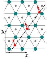
Recent experimental studies have found that magnetic impurities deposited on superconducting monolayer NbSe2 generate coupled Yu-Shiba-Rusinov bound states. In this context, we considered ferromagnetic chains of impurities which induce a Yu-Shiba-Rusinov band and harbor Majorana bound states at the chain edges. We found that these topological phases are stabilized by strong Ising spin-orbit coupling in the monolayer and estimated the conditions under which Majorana phases appear as a function of distance between impurities, impurity spin projection, orientation of chains on the surface of the monolayer, and strength of magnetic exchange energy between impurity and superconductor.
[1] D. Sticleț, C. Morari, Phys. Rev. B | (2019) 100:075420 | doi: 10.1103/physrevb.100.075420
- Institute of Water Chemistry, Technische Universität München, GERMANY;
- BioNanoNet Association (BNN), Graz, AUSTRIA;
- National Institute for Biotechnology, Ben-Gurion University of the Negev, Be’er-Sheva, ISRAEL;
- Department of Life and Environmental Sciences, Horia Hulubei National Institute for R&D in Physics and Nuclear Engineering (IFIN-HH), Bucharest;
- Interdisciplinary Research Institute on Bio-Nano-Sciences, Babes-Bolyai University, Cluj-Napoca;
- Iuliu Hațieganu University of Medicine and Pharmacy, Cluj- Napoca;
- The Institute of Interdisciplinary Research, Alexandru Ioan Cuza University, Iasi;
- National Institute for Research & Development in Chemistry and Petrochemistry ICECHIM, Bucharest;
- The National Institute for Research and Development on Marine Geology and Geo-Ecology – GEOECOMAR, Bucharest;
- National Institute for Laser, Plasma and Radiation Physics (INFLPR), Bucharest;
- International Centre of Biodynamics, Bucharest;
- Department of Physics, University of Craiova.
